In this paper half-contour features are proposed to classify benign and malignant breast tumors with PAS considering the fact that the upper half of the tumor contour is less affected by PAS. Acoustic Shadowing and Enhancement Claim Survey.

Sonographic Evaluation Of Benign And Malignant Breast Masses Iame
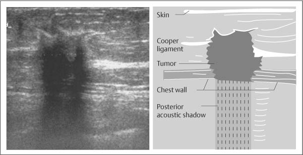
Acoustic shadowing on an ultrasound image is characterized by a signal void behind structures that strongly absorb or reflect ultrasonic waves.
. The results of pathology pending. 2Invasive ductal carcinoma grade 1 in. Hard irregular movable breast masses without pain are typical.
The lesions can be multiple and bilateral 23. A combination of the ultrasonic beams which have propagating characteristics and the tissues in the human body which have acoustic properties give rise to posterior shadowing. As ultrasonic beams propagate through tissues there is a loss of energy by absorption reflection and scattering.
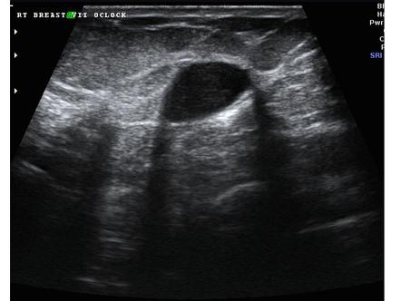
Up to 10 cash back Posterior acoustic shadowing PAS can bias breast tumor segmentation and classification in ultrasound images. However posterior acoustic shadowing caused by a desmoplastic reaction can be found in benign breast neoplasms as well. While breast US has certain advantages over digital mammography it suffers from image artifacts such as posterior acoustic shadowing PAS presence of which often obfuscates lesion margins.
Shadowing may result because of reflection of mo. Posterior acoustic features are described as no posterior acoustic features enhancement white shadowing dark or a combined pattern. This finding is an important one to recognize and distinguish from several other causes of isolated acoustic shadowing.
Up to 20 cash back I had a breast ultrasound with core biopsy. While breast US ha Distinguishing Lesions from Posterior Acoustic Shadowing in Breast Ultrasound via Non-Linear Dimensionality Reduction - IEEE Conference Publication. Posterior acoustic shadowing is a suspicious finding and may be seen in cases of invasive carcinoma postoperative scar complex sclerosing lesion or macrocalcifications and may even be seen in patients with dense breast tissue.
Breast Ultrasound Past. On mammography the lesion usually shows localized increased density in the glandular tissue. The calculi in the kidney and gallbladder cause pronounced posterior acoustic shadowing.
We are developing new feature extraction and classification methods for computerized characterization of posterior acoustic patterns of breast masses into shadowing no pattern or enhancement categories. This mass was categorized as BI-RADS 5. For example fibrosis inside a tumor can block ultrasound from passing deeper causing acoustic shadowing.
Breast sonogram shows 11-cm mass displaying irregular shape angular margins nonparallel orientation echogenic halo hypoechoic echotexture and posterior acoustic shadowing. Posterior acoustic features may or may not be seen when imaging solid masses. While breast US has certain advantages over digital mammography it suffers from image artifacts such as posterior acoustic shadowing PAS presence of which often obfuscates lesion margins.
Adaptive thresholding and disk. An irregular hypoechoic mass with intense posterior acoustic shadowing can be typically seen on US and can mimic breast malignancy Fig. Posterior acoustic shadowing PAS can bias breast tumor segmentation and classification in ultrasound images.
It is wider than tall with macrolobulations no calcifications and posterior acoustic shadowing. Breast ultrasound US in conjunction with digital mammography has come to be regarded as the gold standard for breast cancer diagnosis. Breast ultrasound US in conjunction with digital mammography has come to be regarded as the gold standard for breast cancer diagnosis.
In breast sonography hypoechoic nodules with posterior shadowing can prove to be a major hindrance in the test for malignancy. Posterior acoustic enhancement and shadowing on ultrasound US images are important features used by radiologists for characterization of breast masses. After 5 years of excisional biopsy lesion shows irregular hypoechoic mass with posterior acoustic shadowing on transverse ultrasonography.
The boundary between the mass and the surrounding tissue is described as having an abrupt interface or as containing an echogenic halo a white blurry band surrounding the mass. While breast US has certain advantages over digital mammography it suffers from image artifacts such as posterior acoustic shadowing PAS presence of. The phenomenon of acoustic shadowing sometimes somewhat tautologically called posterior acoustic shadowing on an ultrasound image is characterized by a signal void behind structures that strongly absorb or reflect ultrasonic wavesIt is a form of imaging artifactThis happens most frequently with solid structures as sound conducts most rapidly in.
This loss is displayed in the image as shadowing and is an important sonographic. Ultrasound Artifacts 2. AbstractBreast ultrasound US in conjunction with digital mammography has come to be regarded as the gold standard for breast cancer diagnosis.
In this paper half-contour features are proposed to classify benign and malignant breast tumors with PAS considering the fact that the upper half of the tumor contour is less affected by PAS. It is the posterior acoustic shadowing that is freaking me out. Initially she had left breast lesion that was confirmed as fibrocystic change with stromal fibrosis on excisional biopsy not shown.
However posterior acoustic shadowing caused by the desmoplastic reaction can be found in benign breast neoplasm too. The hepatic cyst demonstrates the opposite phenomenon of posterior acoustic enhancement. Download scientific diagram Transverse ultrasound of the left breast demonstrates an irregular antiparallel mass with posterior acoustic shadowing.
Theultrasound showed a 40 x 24 x 40cm irregular mass with angular margins posterior shadowing and architecturl distort. Upon a screening mammogram and ultrasound they found a 16 oval mass on my right breast. Acoustic Shadowing Although invasive breast cancer is usually identified at sonography as a visible mass a focus of acoustic attenuation or shadowing without a definable mass may be the only feature identified Fig.

Posterior Acoustic Shadowing In Benign Breast Lesions Weinstein 2004 Journal Of Ultrasound In Medicine Wiley Online Library

Mediconotebook Posterior Acoustic Shadowing And Enhancement

Ultrasound Image Of A Breast Cancer With Irregular Borders Angular Download Scientific Diagram

Posterior Acoustic Shadowing In Benign Breast Lesions Weinstein 2004 Journal Of Ultrasound In Medicine Wiley Online Library
Benign And Malignant Characteristics Of Breast Lesions At Ultrasound Radiology Reference Article Radiopaedia Org
2
Basic Principles Radiology Key
![]()
Transverse Ultrasound Of The Left Breast Demonstrates An Irregular Download Scientific Diagram
0 comments
Post a Comment